Head And Neck Muscle Diagram - 3 - Human anatomy for muscle, reproductive, and skeleton.. Obliquus capitis superior also extends from the occiput to c1 while obliquus. Working in pairs on the. Mouse over each are to identify the muscle. Human anatomy diagrams show internal organs, cells, systems, conditions, symptoms and sickness information and/or tips for healthy living. For more anatomy content please follow us and visit our website:
The skull can be further subdivided into. § voluntary somatic muscle § make up the skeletal muscles cardiac striated muscle § involuntary visceral muscle § forms most of the walls of the heart and adjacent parts of muscle attachment sites. This article describes the anatomy of the head and neck of the human body, including the brain, bones, muscles, blood vessels, nerves, glands, nose, mouth, teeth, tongue, and throat. Muscles of head and neck. The neck contains several organs and pathways and is stabilized by muscles, which form the main part of the neck.
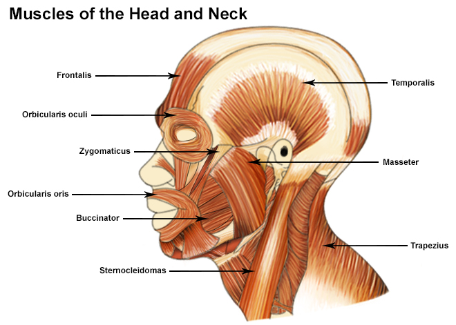
Visual Guide To Cancers Of The Head And Neck from img.webmd.com At the front, the muscles reach from the jawbone to the sternum and clavicle bones, helping us to move our jaw. 9 видео 363 062 просмотра обновлен 5 февр. They are attached to the femur (thighbone), tibia (shinbone), and fibula (calf bone) by fibrous tissues called deep lymphatics of head and neck. Working in pairs on the. The head rests on the top part of the vertebral column, with the skull joining at c1. The neck is the connection between the head and torso. Head and neck muscles diagram. Related posts of muscle of the neck and head.
Learn this topic fast with head and neck muscle anatomy reference charts. Muscles that act on the head and neck. The skull can be further subdivided into. Working in pairs on the. Mouse over each are to identify the muscle. This muscle originates at some of the spinous processes in your thoracic vertebrae (the thoracic vertebrae are the ones where your ribs connect), and it inserts at the transverse processes of the first. Head and neck muscles diagram so many muscles that cause migraines arm neck shoulders and back learn all about it at kenhub. Neck neck muscle anatomy muscle diagram inspirational medical. § voluntary somatic muscle § make up the skeletal muscles cardiac striated muscle § involuntary visceral muscle § forms most of the walls of the heart and adjacent parts of muscle attachment sites. The neck contains several organs and pathways and is stabilized by muscles, which form the main part of the neck. We hope this picture head and neck muscles diagram can help you study and research. The neck muscles move the head in every direction, working in pairs on either side of the body. As a section of the spine, the cervical vertebrae allow movement of the head and thus expand the range of human perception.
Anatomy of the head and neck (ct scan). Head and neck anatomy is important when considering pathology affecting the same area. The splenius capitis and splenius cervicis, which are located in the back of the neck, work to rotate the head. The muscles that affect the knee's movement run along the thigh and calf. For more anatomy content please follow us and visit our website:
Muscle Neck Images Stock Photos Vectors Shutterstock from image.shutterstock.com The muscles that affect the knee's movement run along the thigh and calf. The muscles of the head and neck are all axial. Mouse over each are to identify the muscle. Human anatomy diagrams show internal organs, cells, systems, conditions, symptoms and sickness information and/or tips for healthy living. The head rests on the top part of the vertebral column, with the skull joining at c1. The suboccipital muscles act to rotate the head and extend the neck. At the front, the muscles reach from the jawbone to the sternum and clavicle bones, helping us to move our jaw. Human anatomy for muscle, reproductive, and skeleton.
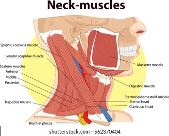
The neck muscles, including the sternocleidomastoid and the trapezius, are responsible for the gross motor movement in the muscular system of the head and neck.
Working in pairs on the. The deep neck muscles (figure 7.18 and table 7.7) include neck flexors, located along the anterior surfaces of the vertebral bodies, and neck extensors, located posteriorly. Rectus capitis posterior major and rectus capitis posterior minor attach the inferior nuchal line of the occiput to the c2 and c1 vertebrae respectively. As a section of the spine, the cervical vertebrae allow movement of the head and thus expand the range of human perception. The suboccipital muscles act to rotate the head and extend the neck. Mouse over each are to identify the muscle. Muscles of the shoulder are a group of muscles surrounding the shoulder joint, which move and provide muscles of the arm and shoulder (labeled diagram). Head and neck anatomy is important when considering pathology affecting the same area. Obliquus capitis superior also extends from the occiput to c1 while obliquus. They are attached to the femur (thighbone), tibia (shinbone), and fibula (calf bone) by fibrous tissues called deep lymphatics of head and neck. This muscle originates at some of the spinous processes in your thoracic vertebrae (the thoracic vertebrae are the ones where your ribs connect), and it inserts at the transverse processes of the first. Audiologist, speech therapist & cochlear implant specialist. In radiology, the 'head and neck' refers to all the anatomical structures in this region excluding the central nervous system, that is, the brain and spinal cord and their associated vascular structures and encasing.
As a section of the spine, the cervical vertebrae allow movement of the head and thus expand the range of human perception. Human muscle system, the muscles of the human body that work the skeletal system, that are under voluntary control, and that are concerned with movement, posture, and balance. Obliquus capitis superior also extends from the occiput to c1 while obliquus. The deep neck muscles (figure 7.18 and table 7.7) include neck flexors, located along the anterior surfaces of the vertebral bodies, and neck extensors, located posteriorly. The next life study seated female figure, shows the upper part of the pectoralis major positioned flat against the rib cage, with very the upper portion of the muscle also helps bend the neck and head backward (extension of the neck).
Seer Training Muscles Of The Head And Neck from training.seer.cancer.gov Muscles of the shoulder are a group of muscles surrounding the shoulder joint, which move and provide muscles of the arm and shoulder (labeled diagram). We hope this picture head and neck muscles diagram can help you study and research. The neck muscles, including the sternocleidomastoid and the trapezius, are responsible for the gross motor movement in the muscular system of the head and neck. Human anatomy for muscle, reproductive, and skeleton. Muscles that act on the head and neck. Many in the neck help to stabilize or move the head. Human anatomy diagrams show internal organs, cells, systems, conditions, symptoms and sickness information and/or tips for healthy living. The head rests on the top part of the vertebral column, with the skull joining at c1.
The skull can be further subdivided into.
The neck muscles, including the sternocleidomastoid and the trapezius, are responsible for the gross motor movement in the muscular system of the head and neck. § voluntary somatic muscle § make up the skeletal muscles cardiac striated muscle § involuntary visceral muscle § forms most of the walls of the heart and adjacent parts of muscle attachment sites. Only two of the more obvious and superficial neck. In radiology, the 'head and neck' refers to all the anatomical structures in this region excluding the central nervous system, that is, the brain and spinal cord and their associated vascular structures and encasing. Anatomy of the head and neck (ct scan). Neck neck muscle anatomy muscle diagram inspirational medical. Discover ideas about muscle diagram. The quizzes below each include 15 multiple choice identification questions related to the muscles of the head and neck. Head and neck anatomy is important when considering pathology affecting the same area. Rotation and lateral flexion of the head are accomplished by lateral and posterior neck muscles. Head and neck muscles provide us functions like expressions, head moving, neck rotating, etc. At the front, the muscles reach from the jawbone to the sternum and clavicle bones, helping us to move our jaw. The splenius capitis and splenius cervicis, which are located in the back of the neck, work to rotate the head.
The neck muscles, including the sternocleidomastoid and the trapezius, are responsible for the gross motor movement in the muscular system of the head and neck neck muscle diagram. Rectus capitis posterior major and rectus capitis posterior minor attach the inferior nuchal line of the occiput to the c2 and c1 vertebrae respectively.


0 Komentar